Bone Gold Radiographs
The following reports are actual Veterinary Case Reports to horse owners and trainers of some of our more interesting clinical presentations. The owners have requested the horses remain totally anonymous.
Report 3: Pedal Bone Fracture Resolution
HORSE: XXXXXXX
DATES OF EXAMINATION: 26TH AUG 21ST OCT 21 & 17TH NOV ‘21
PLACE: XXXXXXXXXX
Radiographs:
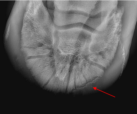
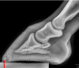
26.08.21: Right Front: Large P3 solar margin fracture evident on medial toe 32.6mm x 4.1mm with approximately 1.4mm separation from parent bone as below left image.
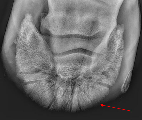
17.11.21: Right Front: Fracture fully resolved.
Recommendations:
Radiographic results as of the 17th of Nov '21 show total resolution of the original fracture.
- XXXXXXXX was reshod today utilising off an alloy shoe with a toe clip - inner circumference seated out to avoid any sole pressure.
- The horse can return to training.
- Continue Bone Gold supplementation at 3 x scoops once per day.
Comments:
- Solar margin Type IV fractures of this magnitude have well documented internationally published healing rates of 7 - 12 months. Full resolution of these fractures is rare as most often the fracture fragments are resorbed. Full resolution in a 3-month period was not anticipated. The horse was on Bone Gold 3 x scoops fed once per day for this period.
Report 4: Pedal Bone Fracture Resolution
HORSE: “XXXX XXXXXX”
MICROCHIP: 9851000121510XX
DATE OF EXAMINATION: 8TH MAR 21 – 6TH JUN 2021
PLACE: XXXXXXXXX FARM
XXXX XXXXXX was referred by Dr XXXXX XXXXX subsequent to a persistent chronic left front lameness over a 7 month period.
Clinical examination on 8th Mar 2021 of XXXX XXXXXX showed a shifting lameness in front predominantly 3/5 L Front. Both front feet were excessively warm to the touch with exaggerated medial and lateral pulses. Palpation with hoof testers found significant discomfort of the left front medial toe
Radiographic Examination:
Radiographs on the 8th Mar 2021 of the left front hoof showed a 7.4cm Type IV solar margin fracture.
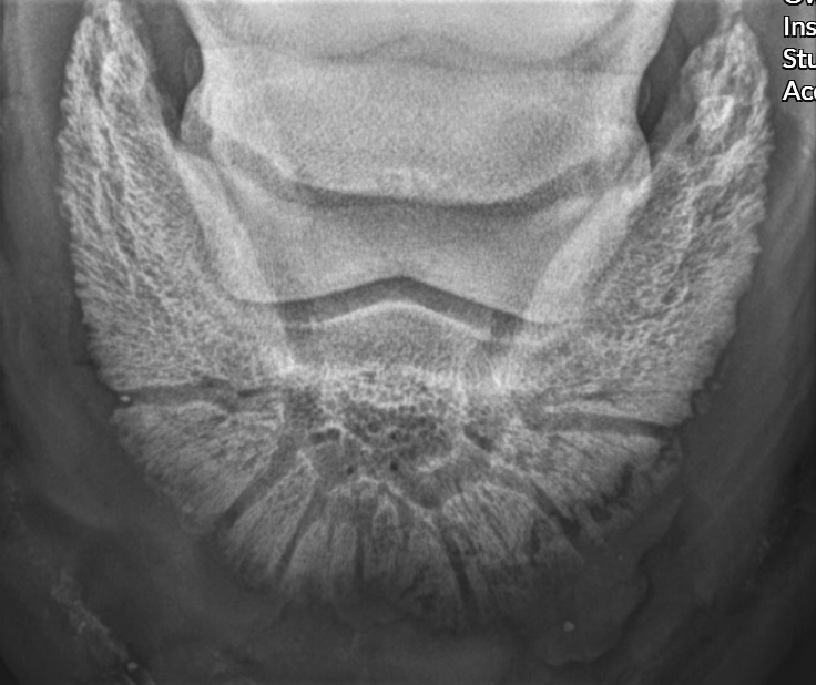
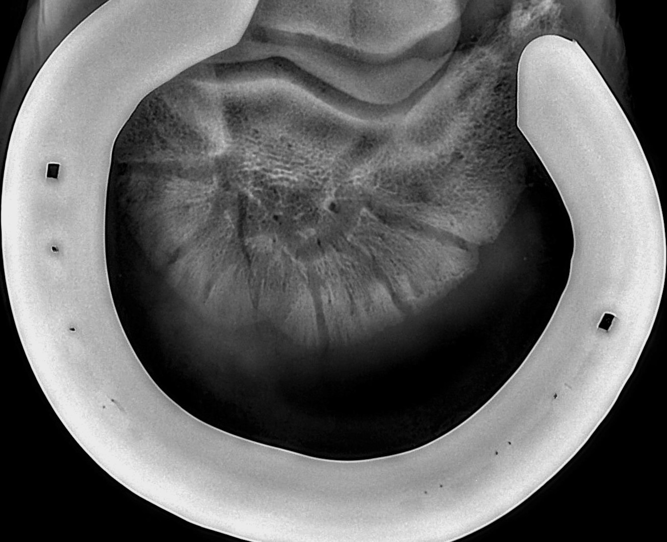
Remedial shoeing was performed and recommendations to re radiograph in 3 months were made to monitor progress. Radiographs on the 12th Jun 2021 clearly identify total resolution of the fracture.
Recommendations:
- Continue to monitor closely and maintain remedial shoeing.
Comments:
- The size and discomfort represented by this particular fracture necessitated a recommendation of a slow return to performance over 8 to 12 months.
- Radiograph taken 14 weeks post initial consultation show total healing of the fracture.
- The Colt has a good prognosis for return to full racing.
- Note that this is an unusually fast recovery.
- The Colt started Bone Gold supplementation on 12th Jun 2021 at 3 x scoops fed once per day, continue daily supplementation at the same rate.
Report 5: Laminitis Resolved
VETERINARY REPORT
HORSE: XXX XXXXXXXX
DATE OF EXAMINATION: 25TH FEB 2021
PLACE: XXXXXXXXX XXXX CANUNGRA
XXX XXXXXXXXX was examined on 25th Feb and found to be 4/5 lame at the walk left front. Examination revealed a deficit in the sole cranial to the tip of the frog discharging serum. Radiographs were taken showing a 4-degree rotation of P3 in the left with associated acute pedal osteitis and minor demineralisation of P3 solar margin with a clear sub solar gas line on lateral view indicating rotational laminitis. The right front shows no radiographic laminitic changes.
The heels were raised with polyurethane and sole packed with the open sole curetted and packed with antibiotics. Post treatment mobility and soundness has improved. The horse will be carefully monitored and kept in box rest with additional radiographs to be retaken in 2 weeks to monitor progress and stabilisation of P3 rotation.
Cause of laminitic incident unknown - possible sequelae to stifle surgery.
Radiographic Examination:
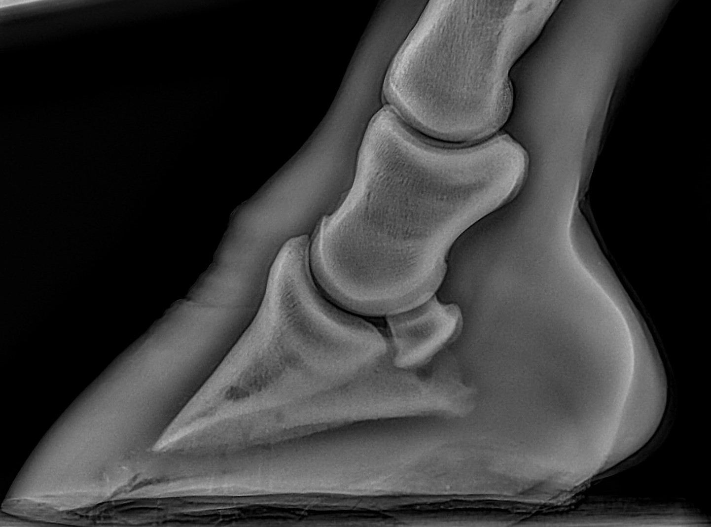
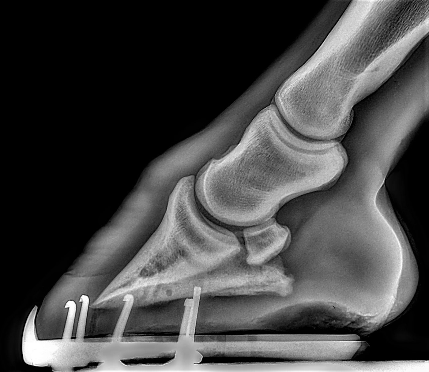
Radiographs on the 25th Feb 21 clearly show a rotated P3 with an air pocket continuous from the sole to the ventral extremity of the Pedal bone and approximately 4mm of sole depth despite ventral capsular rupture.
On clinical examination on 26th May XXX XXXXXXXX presented sound at the trot, sole integrity was excellent and negative to testers.
The ventral sole is beginning to show concavity, wall integrity has improved and approximately 3.12cm of wall growth from the dorsal coronary band was radiographically measured from 25th Feb to 26th May.
Comments:
- The recovery time span for the colt has been remarkable.
- Initial prognosis for return to racing has reverted from poor to good.
- The colt has been on Bone Gold 3 x scoops once per day since initial consultation and should remain on the supplement for 6 months as a prophylactic against potential relapse.
- Continue remedial shoeing with core support for 8 weeks.
Report 6: Multiple Pedal Bone Fractures Resolved
VETERINARY REPORT
HORSE: XXXXXX XXXXX
DATE OF EXAMINATION: 15TH OCT / 28TH DEC 2021
PLACE: XXXXXXXXX FARM
XXXXX XXXXX was presented 15th Oct 21 3/5 lame L Fr with an elevated digital pulse and extremely sensitive to testers around peripheral sole margin.
The colt blocked sound to a low point block to the left front. Of note, both the wall and sole integrity is poor and brittle with solar surface partially convex.
Radiographic Examination:
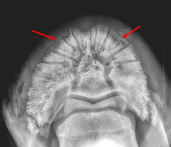
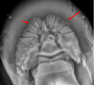
Two large P3 solar margin fractures can be identified on medial and lateral peripheral margin quadrants on the left front. The lateral fracture measures 4.8cm in length and the medial fracture measures 3.6cm.
Recommendations:
The colt was shod with broad aluminium shoes with titanium toe insert seated out to the widest circumference of the sole with side clips pulled medially and laterally. A Castle pad was applied with CS Sole pack and the colt re evaluated on 28th Dec 21.
Comments:
- The horse was sound on re-examination on 28th Dec (10 weeks).
- Radiographs taken on 28th Dec 21 show the sole depth had increased radiographically from 4.1mm to 9.3mm in an 8-week period with no sensitivity to testers.
- Both the medial and lateral solar margin fractures show 90% resolution.
- Continue remedial shoeing with core support for 8 weeks and re radiograph.
- Prognosis for full return to racing is good.
- Of note - resolution of fractures of this severity normally require up to 7 - 12 months to heal vs 10 weeks as in this case.
- Continue Bone Gold 3 x scoops once per day.
Report 7: Hind Cannon Fractures Resolved
VETERINARY REPORT
HORSE: X XXXX XXXXXXX
MICROCHIP: …483
DATES OF EXAMINATION: 14TH APRIL / 17TH MAY 2021 /14TH JUNE
PLACE: XXXXX XXXX CANUNGRA
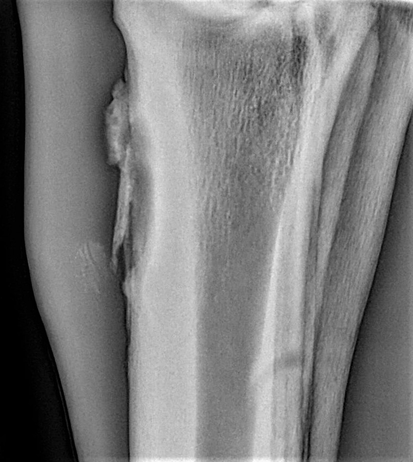
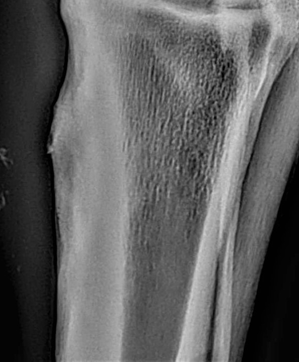
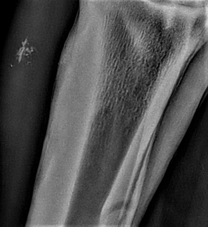
Bilateral hind cannon radiographs of X XXXX XXXXXX were taken on 14th April 21 and again 17th May 21 and 14th June 21. Initial radiographs showed a 32mm longitudinal non displaced left mid cannon saucer fracture extending 6mm into the dorsal cortex and a 44mm longitudinal displaced fracture right mid cannon 5.5mm in depth mid dorsal cortex associated proximally with a 27mm x 6.5mm secondary fracture as below.
Initially, surgical removal was recommended of the R H fracture fragment.
A second series was taken on 17th May and third series on 14th June as above. Both fractures have attached and integrated with the parent bone with minor surface enthesophyte formation and periosteal activity evident.
Recommendations:
Surgery is no longer recommended; however, the colt should be monitored carefully for any swelling on either cannon. Prognosis for full return to racing is good.
Comments:
- Fractures of this nature usually require surgical removal. Resolution of cortical fractures of this size without surgical intervention is not expected.
- Bone Gold supplementation was initiated on 14th April 21 at 3 x scoops/day and should be continued.
Report 8: Post Operative Surgical Keratoma
HORSE: XXXXXX
DATE OF EXAMINATION: 23RD MAR '22
PLACE: XXXXXXXX XXXXXXXXXXX CANUGRA
Full report
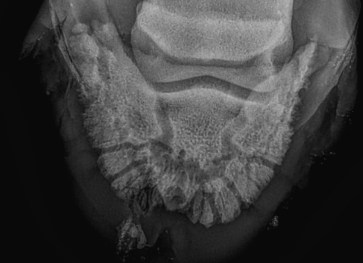
XXXXXX was re radiographed on 23rd Mar, 8 weeks post-surgery 21st Jan.
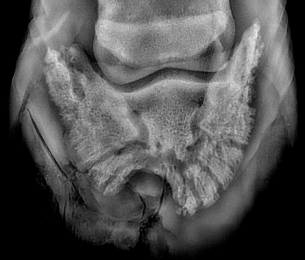
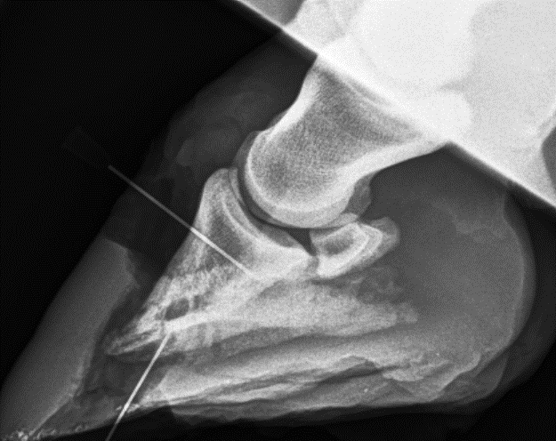
Of note, the horse has been sound since surgery and the surgical sites are clean and healthy. The curetted solar margin radiographically now shows a clearly defined margin on the dorsoventral and lateral views indicating no residual osteomyelitis.
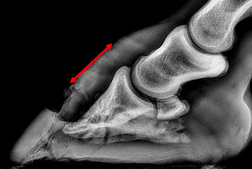
Dorsal hoof wall growth rate 8 weeks post operatively is significant.
Treatment:
XXXXXX was shod all round on 23rd Mar with alloy plates with copper sulphate topically in solar and wall deficits with Sole Pack CS to seal surgical sites.
Dorsal wall measurements radiographically show unusually high growth rates with 38mm of hoof wall growth from the coronary band to the injury.
The left front lateral toe solar deficit was packed with copper sulphate and Sole Pack C
Comments:
International Equine publications indicate expected dorsal hoof wall growth rates to be between 2.5mm - 5mm/30 days. XXXXXX XXXXX has shown nearly 19mm/30 days or approximately 300% higher than expected.
Report 9: Hoof Abscess Drained / Post Operative Surgical Keratoma
HORSE: XXXXXXX
DATE OF EXAMINATION: 23RD MAR / 17TH AUG 22
PLACE: XXXXXXXXXX FARM
Full report
Clinical Examination:
XXXXXX was examined on Aug 17th and found to be sound at the trot. The left front hoof was packed with Solepack CS. The horse has been racing successfully and has been turned out for a spell and re evaluation.
Radiographic Examination:
Radiographs of the left front hoof show considerable consolidation of the ventral sole at the toe with incremental heel height and a positive PA. Radiographic sign of original abscess on 23rd Mar have dissipated.
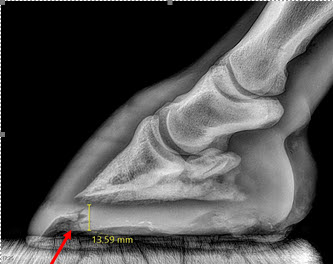
Lateral views show a remarkable 64.2mm of dorsal hoof wall growth in 5 months – nearly 13mm/per month. The surgical resection site is now fully grown out as seen on 17th Aug Lat view on the right.
Comments:
International Equine publications indicate expected hoof growth is approximately 2.5mm – 5mm/30 days. XXXXXXX has radiographically demonstrated growth of 13mm/30days
Recommendations:
- The horse not be left barefoot and should remain shod with quarter clips only all round. Sole pack CS should be applied to the sole of the L Fr and R H as required.
- The prognosis for return for racing is good
- The horse has been on Bone Gold subsequent to surgery in Mar and should remain on 3 x scoops once per day to maintain hoof growth, wall and P3 integrity.
Report 11: Osteomyelitis / Infection Of The Pedal Bone Resolved With Bone Regenerating.
HORSE: XXXXXXXX
MICROCHIP: XXXXXXXXX3311
DATE OF EXAMINATION: 27TH JUL 23
PLACE: XXXXXXXX XXXXXXXXXX
Full report
Clinical Examination:
The horse was sound on presentation at the walk with no significant palmar digital pulse or heat identified in the left front hoof. Surgical site was dry with no evidence of medial/lateral underrunning of dorsal wall.
Radiographic Examination:
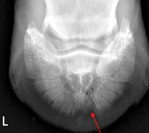
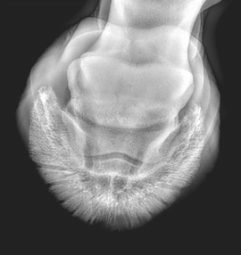
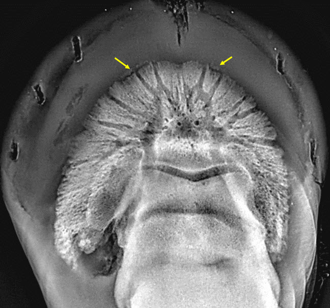
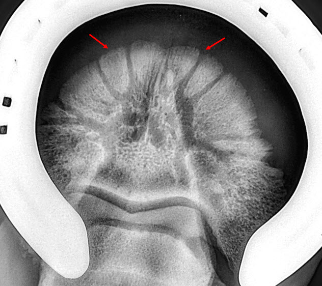
Dorsoventral radiographs show incremental bone density at the surgery site with formation of normal solar margin density and vascular channel formation (red arrows indicating change in bone density and redevelopment).
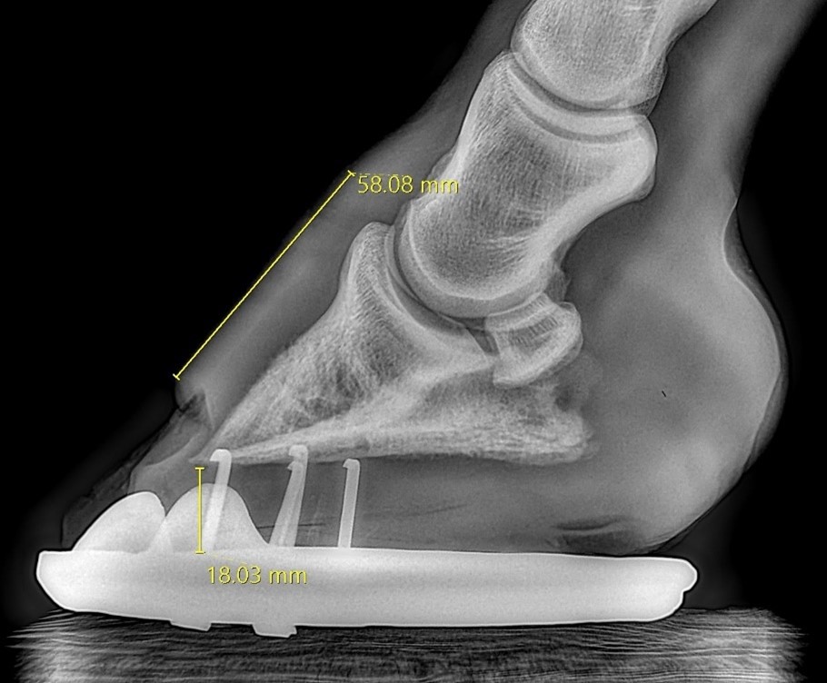
Lateral radiographs show 58mm from coronary band to surgical lesion. Post surgical measurements from coronary band indicate 32mm from coronary to wall surgical site indicating 26mm dorsal wall growth subsequent to surgery.
Comments:
Post surgery the hoof has recovered well with significant dorsal wall growth: 26mm in 45 days, the equivalent of 17mm/30 days (average growth for thoroughbreds is 2.5 - 5mm/30 days)
The solar margin is indicating a return to normal trabecular structure. This rarely occurs and is a good prognostic indicator for full recovery.
Shoeing:
The left front was trimmed and balanced, a steel heart bar applied with sole pack CS. The surgical site was debrided and additionally packed with sole pack CS.
The right front was reshod with a steel heart bar without sole pack.
Recommendations:
- The horse can now be turned out in a small yard without bandaging.
- Continue Bone Gold at 3 x scoops once per day.
Report 12: Hoof Fracture Fragments
HORSE: XXXXXXXXXXX
CHIP: 985100012XXXXXX
DATE OF EXAMINATION: 7TH DEC 23
PLACE: XXXXXXX XXXXXXXXXX
Full report
Clinical Examination:
The horse presented sound at the trot and is in work with no heat or digital pulse evident bilaterally 12 weeks post original radiographs.
Radiographic Examination:
Radiographs of the left and right front feet show total resolution of all fractures despite the 1.5mm previous separation of the fracture fragments from the parent bone.
Comments:
Despite the distraction of the fractures from P3, all fragments have successfully reunited with the P3 margin.
Full resolution of Type IV fractures in a 12 week period is well inside international guides of 6 - 9 months.
Recommendations:
Continue Bone Gold supplementation at 3 x scoops per day while the horse is in work and ensure shoes are well seated out.
